RESEARCH AT NOTRE DAME | COLORECTAL CANCER
A novel three-dimensional cell culture technique is making it possible for researchers to study tumors and evaluate potential cancer therapeutics quicker and more efficiently.
Preclinical cancer research is usually carried out in conventional two-dimensional cell cultures or in animal models. A novel 3D tumor cell culture combines the advantages of both of these approaches by mimicking tumors in vitro more accurately than a traditional 2D approach. Also, it eliminates the need for animal experiments, which are usually costly, time consuming, and involve numerous ethical considerations.

Dr. Amanda Hummon, a Huisking Foundation, Inc. Associate Professor in the Department of Chemistry and Biochemistry at the University of Notre Dame and a member of the Harper Cancer Research Institute, is using the 3D tumor model to study new approaches to combating colorectal cancer, the third most common cancer type.
“One of the great benefits of the 3D tumor model is that potential drugs can be studied on human cancer cells, but the experiments do not need to involve any patients," says Hummon.
Several hundred tumors can be grown in cell culture at a time, creating a so-called high-throughput screening system, where numerous drugs and drug combinations can be studied simultaneously. This model can also be used to examine how various compounds penetrate a tumor—an essential factor for eliminating cancer cells throughout the entire tumor mass, yet something that is not possible to evaluate in a conventional 2D cell culture.
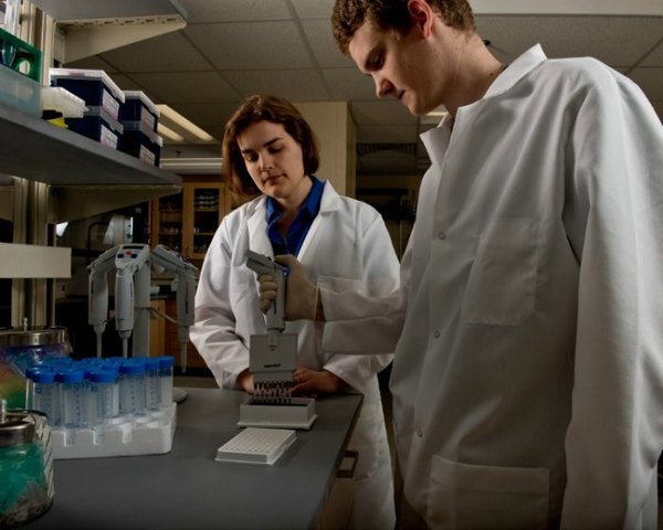
The 3D tumor model can be also utilized to look into drug metabolism and study a wider variety of therapeutic approaches. Gabe LaBonia, a second-year graduate student in Dr Hummon’s laboratory, uses imaging mass spectrometry to look at how different compounds are metabolized by a tumor. The results of his work were very well received at the Pittsburgh Conference on Analytical Chemistry and Applied Spectroscopy in March of 2016.

Another member of the Hummon lab, a third-year graduate student Monica Schroll, is using the 3D tumor model to evaluate how tumors respond to deprivation of glucose and growth factors. Cancer cells usually have a higher metabolic rate than healthy cells, making them particularly sensitive to such “caloric restriction.” Upon glucose or growth factor deprivation, cancer cells enter autophagy, a state where they consume their own organelles, using them as an energy source. The next step in her research involves assessing whether caloric restriction can improve the response to chemotherapy. Monica noted that if this approach proves successful in a tumor model, it would be easy to implement in a clinical trial, where patients would have to be on a restricted-calorie diet for 72 hours prior to receiving chemotherapy.
While Dr. Hummon’s laboratory is focusing on colorectal cancer, the 3D tumor model can be used to study most epithelial cancers. She is collaborating with Dr. Sharon Stack, also a professor in the Department of Chemistry and Biochemistry at Notre Dame and Director of Harper Cancer Research Institute, on using this model to study ovarian cancer.
Originally published by Jenna Bilinski at harpercancer.nd.edu on May 11, 2016.
