
Preparation of fluorescent Au–SiO2 core–shell nanoparticles and nanorods with tunable silica shell thickness and surface modification for immunotargeting
Prakash D. Nallathamby, Juliane Hopf, Lisa E. Irimata, Tracie L. McGinnity, Ryan K. Roeder, Journal of Materials Chemistry B, 2016, 4, 54185428 DOI: 10.1039/C6TB01659F
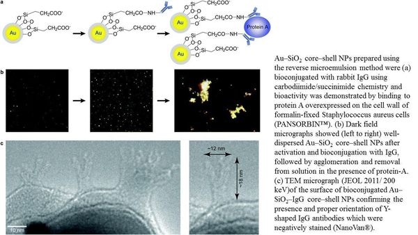
An antibody is a Y shaped protein with high target accuracy. The two branches of the Y shape are always responsible for target specificity of the antibody. For antibody based platform technologies, it’s absolutely imperative that the antibody is standing upright on the surface of the platform. Our study is groundbreaking because the presence and proper orientation of Y shaped antibodies on nanoparticles were directly imaged for the first time in TEM micrographs (JEOL 2011/ 200keV), with high resolution at the nanometer scale, after negative staining. Measured antibody dimensions of ∼18 × 12 nm concur well with unit cell dimensions previously available only from tedious X-ray crystallography measurements. TEM results conclusively demonstrated our ability to correctly orient the antigen binding sites of the antibodies away from the surface of the nanoparticles.
Our antibody imaging technique will be crucial for quality control of immune-targeting antigens in therapeutic, diagnostic imaging, and bio-sensing applications. Moreover, the TEM methodology used in this study is readily transferable to other Y shaped antibodies utilized in the treatment of diseases such as breast cancers, Alzheimer’s, Ebola, and Zika. Our publication underscores the integral and ever expanding role that will be played by electron microscopy in today’s nanotechnology based treatment regimens.
Originally published by at imaging.nd.edu on November 15, 2017.