
On chip three-dimensional tissue histology for microbiopsies
C. Narciso, K. R. Cowdrick, V. Zellmer, T. BritoRobinson, P. Brodskiy, D. J. Hoelzle, S. Zhang, and J. J. Zartman, Biomicrofluidics, 2016, 10, 21101.
We report a novel approach that generates a high resolution, three dimensional (3D) fluorescent staining atlas of tissue microbiopsies in a microfluidic device without destroying the tissue.
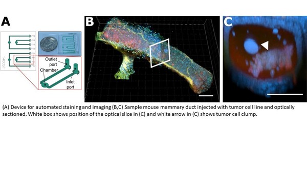
We demonstrate that this method preserves tissue architecture for multiple murine organs by comparing traditional 2D slices to an optically sectioned 3D H&Emimic. The H&Emimic slices show a close qualitative match to traditional H&E. The 3D spatial and molecular information obtainable from this method significantly increases the amount of data available for evaluating both tissue morphology and specific biomarkers in a wide range of both research and clinically driven applications and is amenable to automation.
Originally published by at imaging.nd.edu on November 14, 2017.